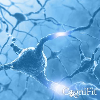
What are neurons? They are tiny cells that are in charge of participating in the functions related to the nervous system. In our brain, there are millions of neurons, scientists calculate that we have about 80 million when we are born. As we grow, this number decreases. After 80 years-old, we will have lost 30% of our neurons. Throughout our life, we constantly lose and regenerate them. By our neurons' regeneration process, new connections are made which produces a process called neurogenesis. This process allows for the birth of new brain cells through a person's life.

Neurons
Reasons why they can live longer and better
Neurocognitive program to train your cognitive abilities.
Online platform where you can train your capabilities and strengthen the weakest ones.
See your results after a few sessions. Sign up and try it!
People do things daily that cause neural deterioration and, as such, cognitive deterioration. These actions, like drinking, smoking, not eating or sleeping well, or stress causes these brain cells to deteriorate more rapidly.
Most people have heard the phrase “use it or lose it”, which is generally applied to physical exercise, but in our neurons' case the same principal can be applied. Here you will see a few reasons why it is necessary to keep your brain cells active.
- Active brain cells receive more blood.
Scientists know that the active areas of the brain use more energy, and thus use more oxygen and glucose. This way, more blood is brought to these areas with the intention of satisfying the active neurons' demands. As you activate your brain, blood runs to the working brain cells. MRI images are used to understand blood flow in the brain. These images have shown that our brain cells are very dependent on the oxygen supply. The more we use our brain and activate them, the more blood supply they receive. On the other hand, an inactive brain cell receives less and less blood until it ultimately dies.
- Active brain cells have more connections with other brain cells.
Each brain cell is connected to its surroundings through rapid fire electrical pulses. Active brain cells tend to produce dendrites, which are like small arms that extend outwards to connect with other cells. One single cell can have up to 30,000 connections. As a result, it becomes a highly active part of the neuronal network. The larger the cell's neuronal network is, the higher the possibility of being activated and surviving.
- Active brain cells produce more “maintenance” substances'.
The Nerve Growth Factor (NGF) is a protein that is produced in your body in the target cells. This protein binds the neurons, marking them as active, differentiated, and responsive. The more often you challenge, exercise, and active your brain, the more NGF is produced.
- Active brain cells stimulate the migration of the beneficial cells from the brainstem.
Recent studies have shown that the new brain cells are generated in a specific area of the brain, called the hippocampus. These brain cells can migrate to the areas of the brain that need it most. For example, they would migrate to a certain area after a brain injury. These migrating cells are able to imitate the actions of the surrounding cells, allowing for a partial restoration of the damaged area.
Structure of a neuron
The neuron is made up of a structure whose main parts are the nucleus, the cell body, and the dendrites. There are many connections between them due to the axons, or small branches. The axons help to create networks whose functions is to transmit messages from neuron to neuron. This process is called synapsis, which is the binding of the axons by electrical charges at a rate of 0.001 seconds, which can occur about 500 times per second.

1. Nucleus
It is the central part of the neuron. It is located in the cell body, and is in charge of producing energy for the cells' functions.
2. Dendrites
Dendrites are the “arms of the neuron”, they form branch extensions that come out of different parts of the neuron. In other words, it is the cell body. The cell usually has many branches, and the size depends of the neuron's function and where it is situated. Its main function is the reception of stimuli from other neurons.
3. Cell body
This is the part of the neuron that includes the nucleus. It is in this space where most of the neuron molecules are synthesized or generated and the most important activities are carried out to maintain life and take care of the functions of the nerve cell.
4. Glial cells
Neurons are specialized cells that by themselves they cannot perform all the nutrition and support functions necessary for their survival. For this reason, the neuron surrounds itself with other cells that perform these functions: Astrocyte mainly responsible for nourishing, cleaning and supporting neurons; Oligodendrocyte mainly responsible for covering the axons of the central nervous system with myelin, although it also performs functions of support and union; Microglia mainly responsible for the immune response, as well as removal of waste and maintenance of neuron homeostasis; Schwann cell responsible for covering the axons of the peripheral nervous system with myelin, as shown in the picture; Ependymocyte responsible for covering the cerebral ventricles and part of the spinal cord.
5. Myelin
Myelin is a material composed of proteins and lipids. It is found forming sheaths around neuronal axons, which allows them to be protected, isolated and transmit up to 100 times more efficiently the potential for action. In the central nervous system, myelin is produced by oligodendrocytes, while in the peripheral nervous system, it is produced by Schwann cells.
6. Axon terminal
Axon terminals, or synaptic boutons, are found at the end of the axon of the neuron, divided into terminals whose function is to link other neurons and create a synapse. The brain's neurotransmitters are stored in the synaptic boutons in small areas called synaptic vesicles. The transmission of these vesicles from the terminal buttons of one neuron to the dendrites of another neuron is what is known as synapses.
7. Node of Ranvier
The Node of Ranvier is a gap or space between each myelin sheath of the axon extensions. The space between each sheath is just enough and is necessary to optimize impulse transmission and ensure that it does not get lost. This is what is known as nerve impulse jump conduction. The main function of the Node of Ranvier is to facilitate movement and optimize energy consumption.
8. Axon
The axon is another main part of the neuron. It is a fine and long nerve fiber that is responsible for transmitting the electric signals between these brain cells. As was previously mentioned, axons have nerve endings wrapped in myelin sheaths that are responsible for transmitting electrical signals from the soma of the neuron to the terminal buttons.
References
James Siberski, Evelyn Shatil, Carol Siberski, Margie Eckroth-Bucher, Aubrey French, Sara Horton, Rachel F. Loefflad, Phillip Rouse. Computer-Based Cognitive Training for Individuals With Intellectual and Developmental Disabilities: Pilot Study - The American Journal of Alzheimer’s Disease & Other Dementias 2014; doi: 10.1177/1533317514539376
Preiss M, Shatil E, Cermakova R, Cimermannova D, Flesher I (2013) Personalized cognitive training in unipolar and bipolar disorder: a study of cognitive functioning. Frontiers in Human Neuroscience doi: 10.3389/fnhum.2013.00108.
Shatil E (2013). Does combined cognitive training and physical activity training enhance cognitive abilities more than either alone? A four-condition randomized controlled trial among healthy older adults. Front. Aging Neurosci. 5:8. doi: 10.3389/fnagi.2013.00008
Peretz C, Korczyn AD, Shatil E, Aharonson V, Birnboim S, Giladi N. - Computer-Based, Personalized Cognitive Training versus Classical Computer Games: A Randomized Double-Blind Prospective Trial of Cognitive Stimulation - Neuroepidemiology 2011; 36:91-9.
Evelyn Shatil, Jaroslava Mikulecká, Francesco Bellotti, Vladimír Burěs - Novel Television-Based Cognitive Training Improves Working Memory and Executive Function - PLoS ONE July 03, 2014. 10.1371/journal.pone.0101472
Korczyn AD, Peretz C, Aharonson V, et al. - Computer based cognitive training with CogniFit improved cognitive performance above the effect of classic computer games: prospective, randomized, double blind intervention study in the elderly. Alzheimer's & Dementia: The Journal of the Alzheimer's Association 2007; 3(3):S171.
Shatil E, Korczyn AD, Peretzc C, et al. - Improving cognitive performance in elderly subjects using computerized cognitive training - Alzheimer's & Dementia: The Journal of the Alzheimer's Association 2008; 4(4):T492.


